
Sinus Lift Procedures – Case study Ι
BEFORE TREATMENT
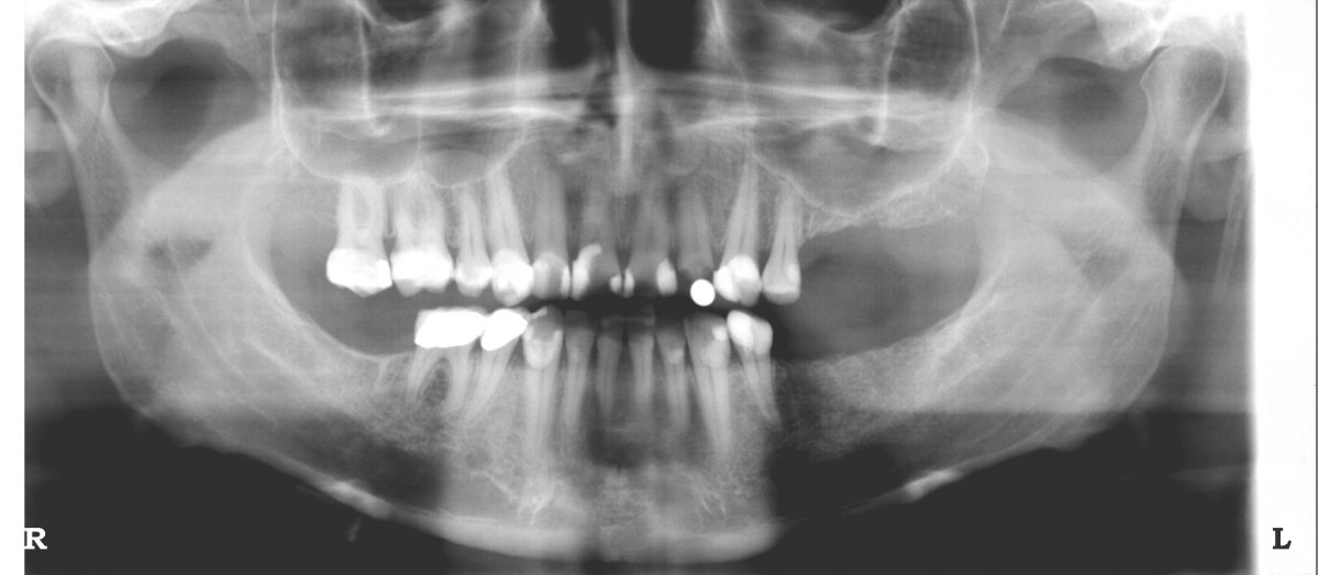
Initial xray. A significant vertical bone loss can be seen on the upper left quadrant that renders an implant placement impossible.

Initial xray. Red color designates the area of the sinus that going toe be grafted with bone in order to increase the available bone height.
AFTER IMPLANT PLACEMENT
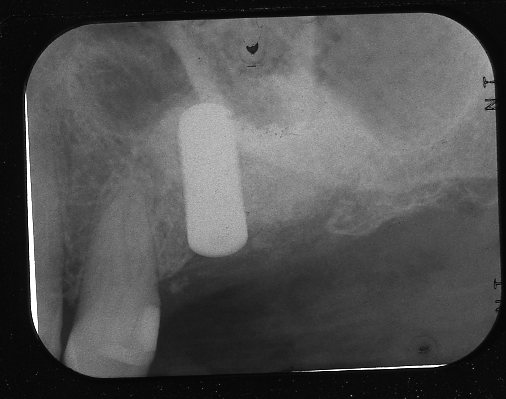
Final xray following implant placement. A sizable implant was placed (4.8mm x 12mm) and it is fully surrounded by bone as seen on the xray. The implant placement and the subsequent restoration of the area with an implant supported porcelain crown were made possible with the sinus lift procedure.
FOLLOWING SINUS LIFT PROCEDURE

Xray taken following the sinus lift procedure. Part of the sinus has been filled with bone graft. The grafted area is more radioopaque (appears more white on the xray due to the presence of bone graft).

Same xray as previously. Blue color represents the grafted area. Due to the significant increase in bone height an implant placement is now possible.