Canine Uncovery – Case study Ι
BEFORE TREATMENT

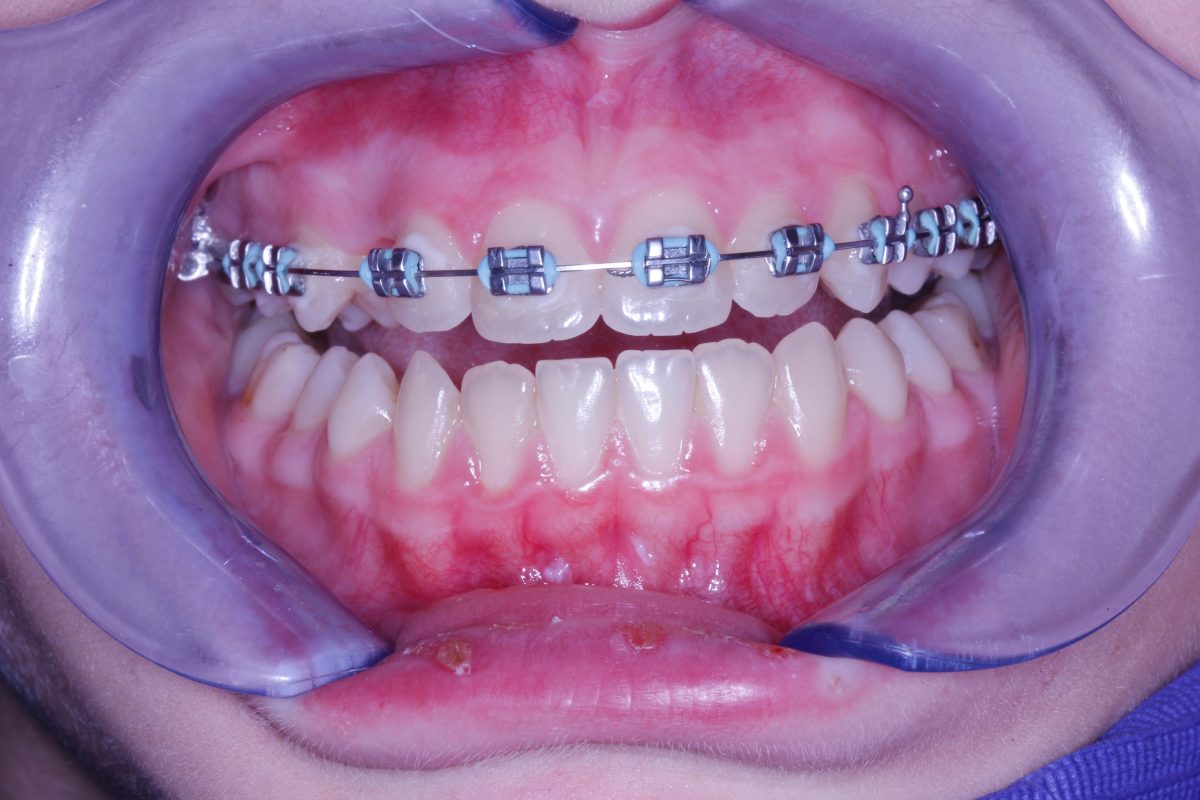
Clinical picture of a case of an impacted upper right canine on a teenage girl (frontal view). The young patient is already undergoing orthodontic treatment by an orthodontic colleague.
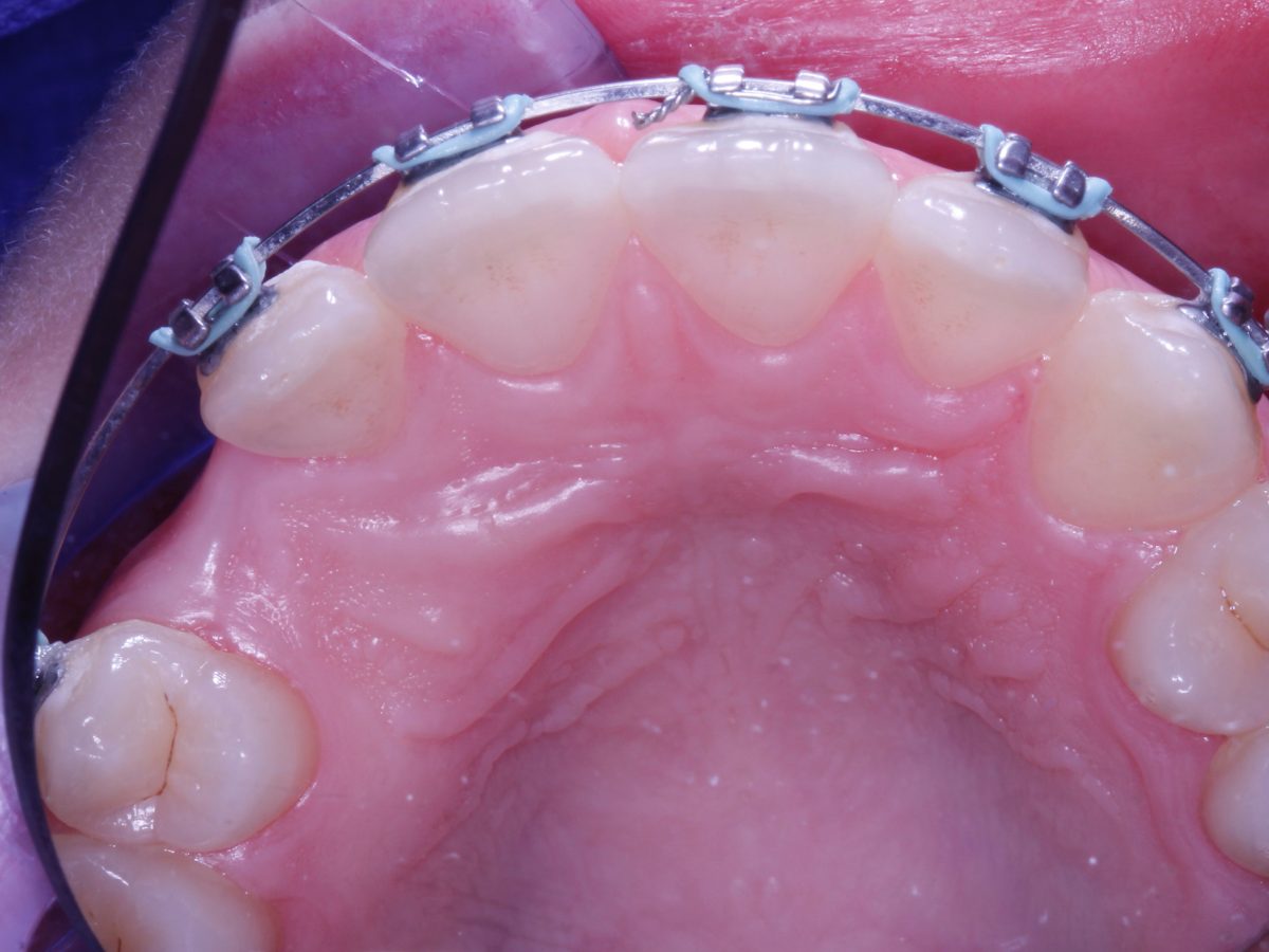
Clinical picture of the same case (palatal/occlusal view). The canine is located under the gums and inside the palatal bone.
TREATMENT
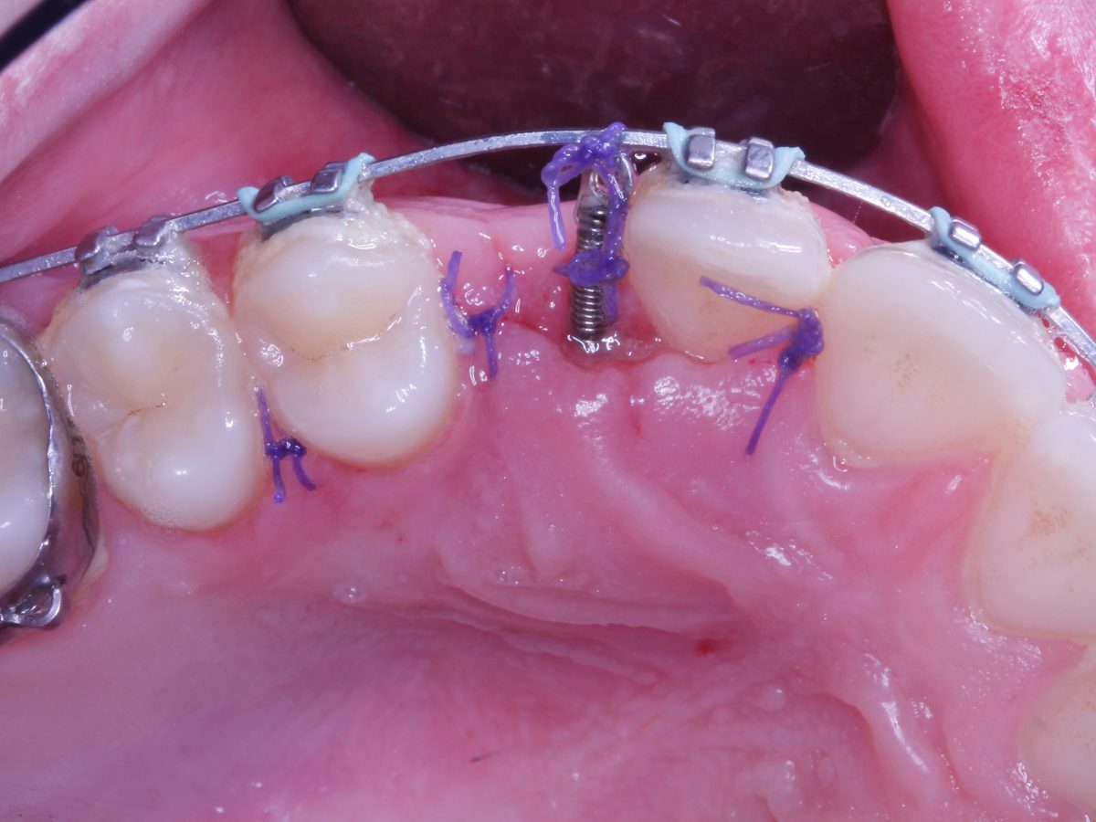
Clinical picture of surgical site. The crown of the canine has been exposed following elevation of the gums and removing the overlying bone.
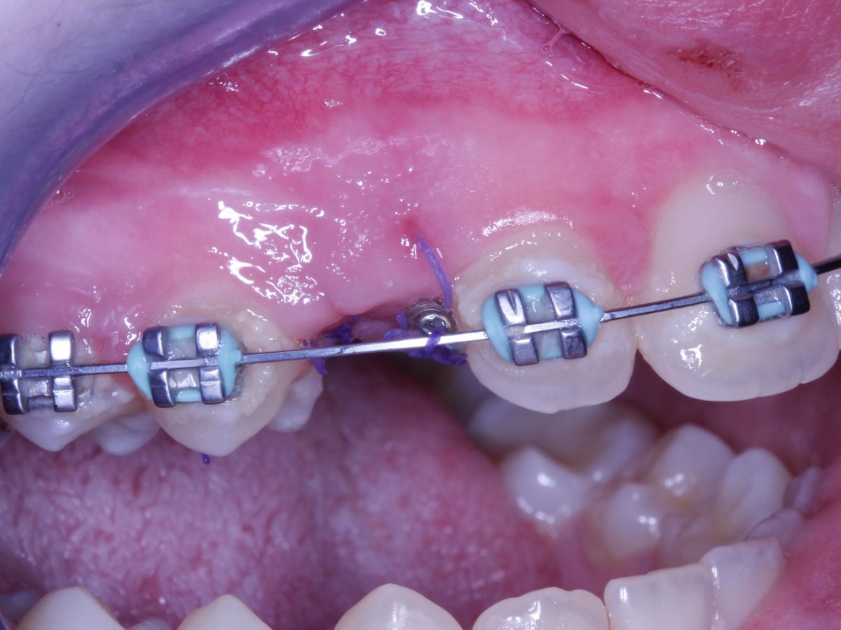
Clinical pictures following the bonding of the orthodontic bracket that will “pull” the tooth and align it correctly with the rest of the teeth. Clean surgical area and integrity of the gums can be seen following careful suturing.
