
Sinus Lift Procedures – Case study ΙI
BEFORE TREATMENT
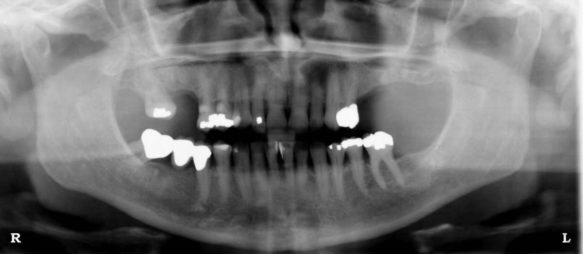
Panoramic xray. A vertical bone deficiency can be seen on the upper left quadrant. No implant placement is possible at this stage.
AFTER SINUS LIFT PROCEDURE

Panoramic xray after the sinus lift procedure. A significan increase in the available bone height is now visible due to the presence of bone graft. Two implants of adequate size may now be placed in the area.
IMPLANT PLACEMENT
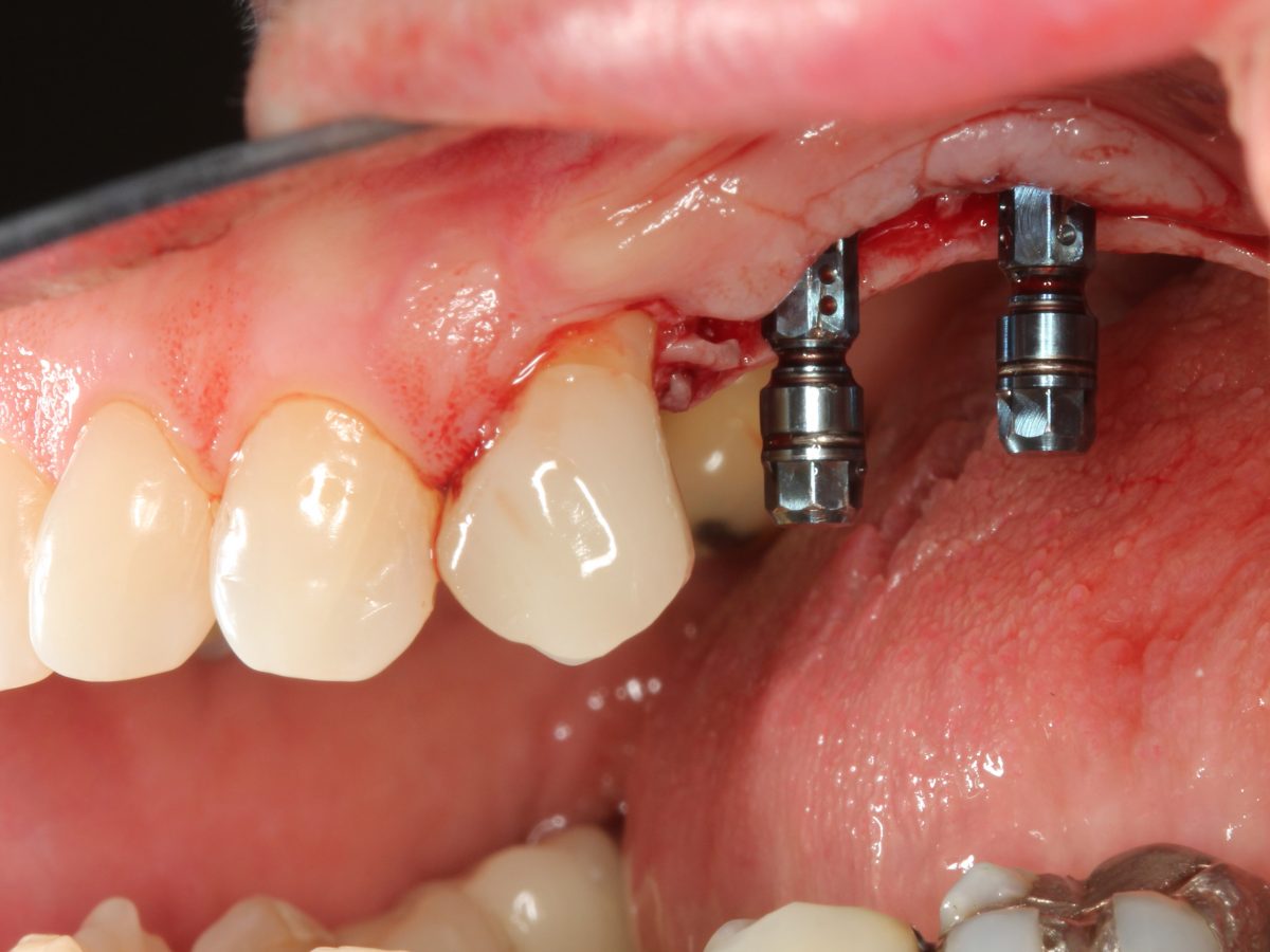
Clinical picture during implant placement. The blue implant carriers confirm the precise parallelism of the implants as well as the ideal positioning of their placement.
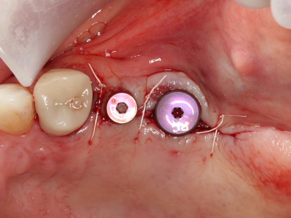
Clinical picture following the placement of healing abutments and suturing. The ideal positioning of the implants in the horizontal plane can be seen as well as the careful handling of the surrounding gingival tissues (gums).
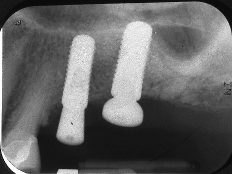
Final xray. The sinus lift procedure enabled the placement of two implants of significant size, which translates into increased bone support for the forthcoming porcelain crowns