
Vertical and Horizontal Bone Augmentation – Case study Ι
In this particular case a guided bone regeneration (GBR) procedure was performed with the use of titanium mesh and a combination of autogenous and allogenic bone graft.
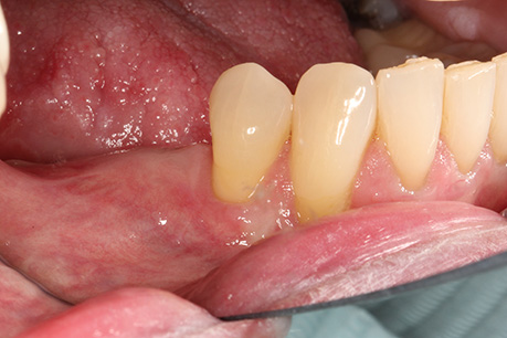
BEFORE TREATMENT
Initial clinical picture. Significant vertical bone loss is evident on the right posterior segment of the lower jaw. This bone loss renders an implant placement problematic.
A cone beam computerized tomography was requested in order to study the case and visualize the bone loss in all 3 dimensions.
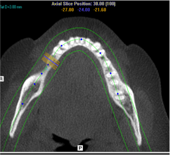
Horizontal ct scan in in which a reestablishment of proper bone thickness following the augmentation procedure can be seen.
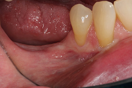
AFTER TREATMENT

Clinical case following the bone augmentation. Complete restoration of the original bone deficiency in vertical dimension, which now enables the placement of dental implants in order to restore the missing teeth.
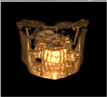
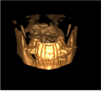
Post bone augmentation 3D Scan. Increase of bone both in horizontal as well as in vertical dimension is evident (increase in height and thickness of bone)
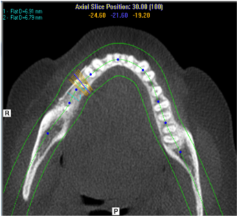
Horizontal ct scanin in which a reestablishment of proper bone thickness following the augmentation procedure can be seen.